
Fig. 3.
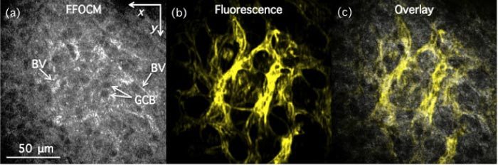
x-y (lateral) images of fixed mouse retina with labeled endothelial cells as acquired with FFOCM (a) and structured illumination fluorescence microscopy (b) in ganglion cell layer. (c) Overlay of (a) and (b). GCB—Ganglion cell bodies; BV—blood vessels.