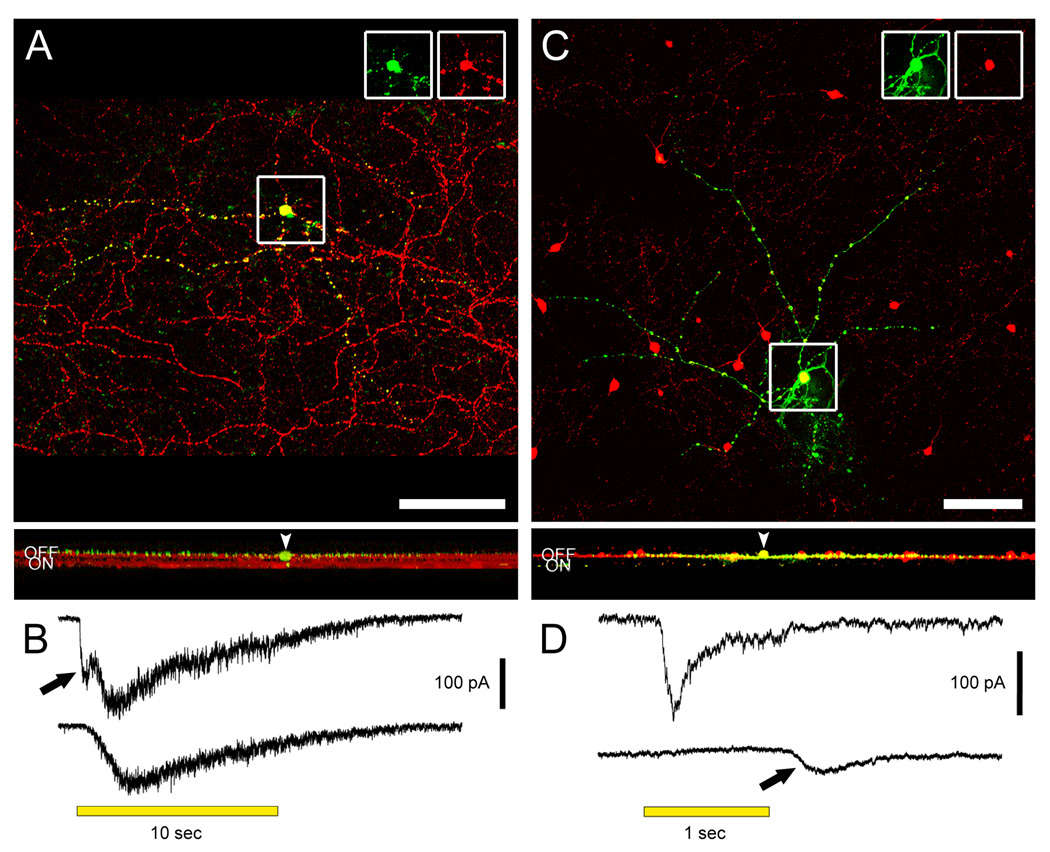
Figure 1. M1 intrinsically photosensitive ganglion cells and dopaminergic amacrine (DA) cells stratify in the OFF sublamina of the IPL, but receive physiological ON channel excitatory inputs.
A. A displaced M1 cell filled with Lucifer Yellow (green) during whole-cell recording and later immunostained against melanopsin (red). Top panel shows the recorded cell and the plexus of melanopsin-positive M1 dendrites in a confocal stack projection of the outer IPL as seen in the whole-mount. Insets at top separate the signals and confirm that the Lucifer Yellow-filled cell expressed melanopsin. Scale bar 100 µm. Bottom panel is a virtual vertical view obtained by 90 degree x-axis rotation and projection of the entire confocal stack. Two bands can be seen in the red channel, corresponding to the level of dendritic stratification levels of M1 cells (upper band; OFF sublamina) and M2 cells (lower band; ON sublamina). Note that all dendrites of the filled M1 cell are restricted to the OFF sublamina. Arrowhead points to the cell body, displaced to the INL. B. Voltage-clamp recordings from the displaced M1 cell in A revealing an ON bipolar cell-driven light response. The recordings were made under conditions that minimized amacrine cell input (see text). Top trace: The control light response consisted of a fast inward current (arrow) that was followed by a slower one. Bottom trace: L-AP4 selectively eliminated the fast component, indicating its generation by ON bipolar cells. C. A dopaminergic amacrine cell expressing RFP (red; TH∷RFP retina) and filled with Alexa488 (green) during whole-cell recording. Top panel shows the cell in the whole-mount view, in the collapsed confocal stack. Insets as for A. Scale bar 100 µm. Bottom panel shows the projected x-axis rotation of the same confocal stack, imaged from the GCL throughout the INL. The red RFP band corresponds to the main dendritic plexus of the dopaminergic cells (outer OFF sublamina). All processes of the filled DA cell are restricted to this sublamina. Very faint green specks lower in the IPL are histological artifacts not traceable to the dye-filled cell. Arrowhead indicates the cell body in the INL. A magenta-green version of the photomicrographs in this figure is available online as a supplemental figure. D. Voltage-clamp recordings from the DA cell in C demonstrating an ON bipolar-mediated light response. Top trace: Under conditions that minimized inhibitory input (see text), an inward current was observed at light onset. Bottom trace: Application of L-AP4 abolished the inward current at light onset, confirming that it was mediated by ON bipolar cells. In addition, a smaller inward current at light off appeared (arrow), which presumably reflects an input from OFF bipolar cells. All light stimuli were −1 log I.

