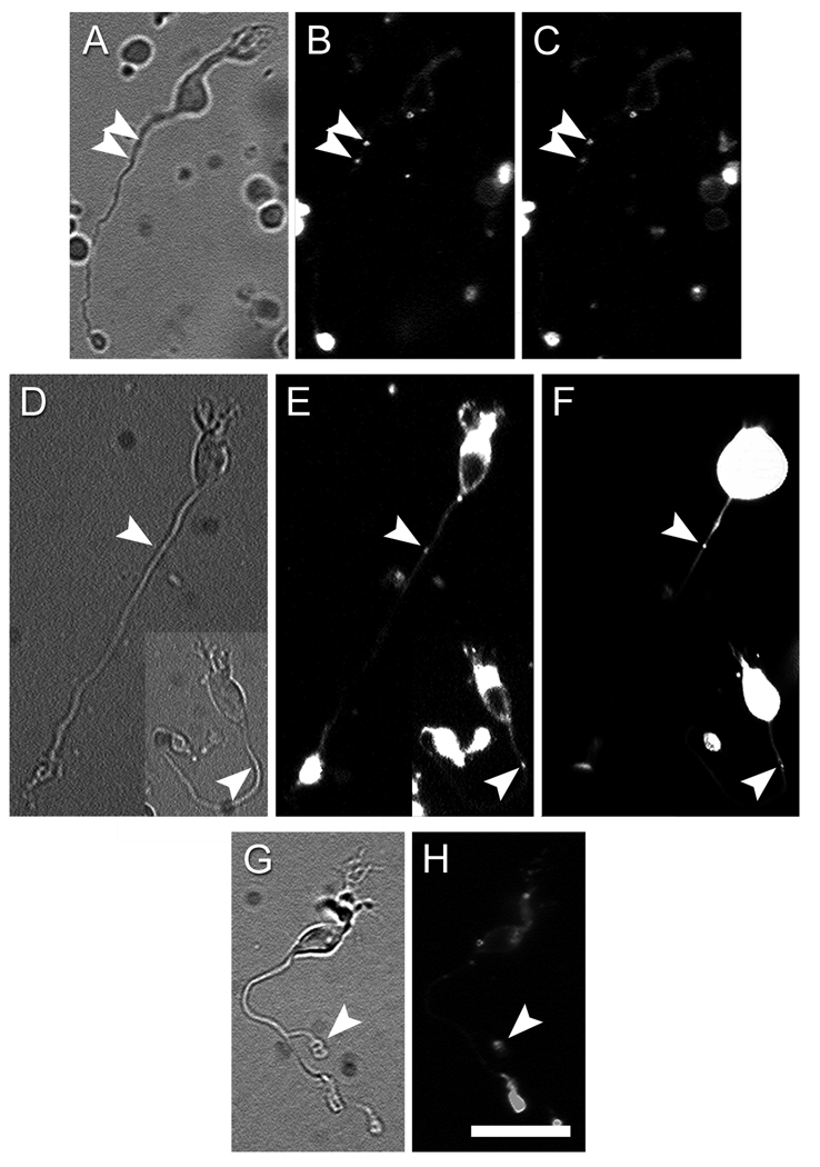
Figure 6. ON bipolar cells can uptake and release vesicles at ectopic synaptic sites.
A – C. Depolarization triggered release of the styryl dye FM4-64 from a dissociated ON bipolar cell. A: Infrared image of the cell. B: FM4-64 staining in control Ringer with low K+ and Ca2+. Arrowheads indicate two puncta of FM4-64 staining in the proximal axon. C: FM4-64 staining of the same cell after 15-min incubation in a high-K+, high-Ca2+ solution. Both puncta are noticeably dimmer when compared with those in the B. D – F. En passant vesicle cycling sites contain synaptic ribbons. In these two ON bipolar cells, en passant FM4-64 staining (arrowheads in E) colocalized with a RIBEYE-binding fluorescent peptide (arrowheads in F). G – H. ON bipolar cell with an ectopic axon terminal (arrowhead in G), which stained for FM4-64 (arrowhead in H). Scale bar = 20 µm.

