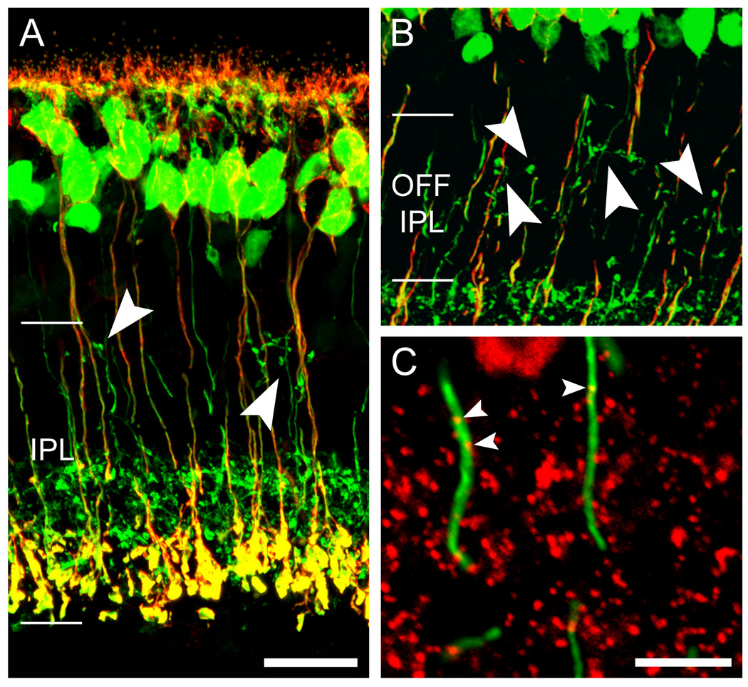
Figure 7. Rod bipolar cells do not appear to contribute to the ectopic ON bipolar output in the OFF IPL.
A & B. Double immunofluorescence as seen in projected confocal stacks of vertical retinal sections showing all ON bipolar cells (green; Grm6-EGFP retina) and rod bipolar cells, marked by PKCα immunostaining (red). Ectopic ON bipolar cell terminals (arrowheads) never colocalize with the rod bipolar cell marker. Scale bar 20 µm. C. High magnification of a wild-type retina double stained against PKCα (green) and CtBP2 (red). Arrowheads point to en passant ribbons in the vicinity of the INL-IPL border. Scale bar 5 µm. A magenta-green version of this figure is available online as a supplemental figure.

