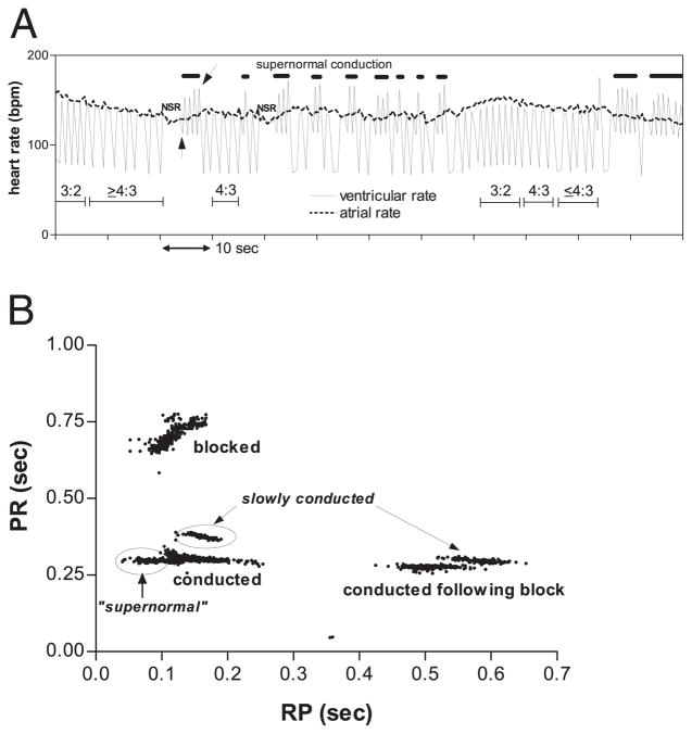
Figure 2. Fetal Heart Rate Patterns and AV Conduction Curves in Supernormal Conduction.
(A) Ventricular and atrial heart rate tracing from last session of fetus #25, showing heart rate pattern changes associated with rhythm transitions. Wenckebach periods, indicated by the thin horizontal bars with conduction ratios above the bars, result in alternation of instantaneous ventricular heart rate between the atrial rate and half the atrial rate. A second pattern of alternation (thick horizontal lines) is due to beat-to-beat alternation of RP and PR, compatible with “supernormal” conduction. The RP and PR interval changes show a paradoxical positive correlation (Fig. 1D), which results in prominent beat-to-beat RR oscillations distinct from those due to Wenckebach periodicity. Usually, this pattern is immediately preceded by several or more beats of 1:1 sinus rhythm with constant RP/PR. The episodes initiate with a long RR interval (upward pointing arrow) and terminate with a short RR interval (slanted arrow) followed by a very long RR interval due to block of the following beat, as exemplified in the rhythm strip in Figure 1D. (B) Scatter plot of PR versus RP for each atrial beat in a 10-min recording during the last session of fetus #25. Atrial beats with RP >170 ms were always conducted, and the probability of conduction varied inversely with RP for 100 ms <RP <170 ms. During episodes of PR alternation (Fig. 1D); however, the data points formed 2 distinct clusters, enclosed by the ovals. One cluster consisted of beats conducted with long PR (slowly conducted); the other consisted of beats that conducted with short RP (“supernormal”). During episodes of PR alternation, beats with RP <100 ms were always conducted. Notice that in the ranges 100 ms <RP <250 ms and 450 ms <RP <650 ms, the conducted beats exhibited 2 distinct PR levels, suggesting the existence of 2 atrioventricular (AV) nodal pathways.

