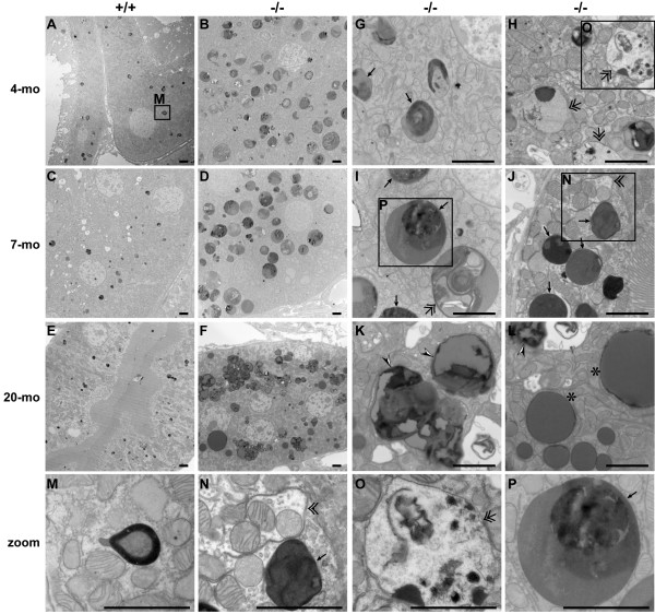
Figure 7.
Electron microscopy analysis of kidneys from LRRK2-/- mice. EM analysis shows the presence of many electron-dense but heterogeneous autophagic vacuoles (autophagosomes (double arrows), autolysosomes (single arrows)) at 4 months of age (A, B, G, H), striking accumulation of giant electron-dense autolysosomes-like structures (single arrows) at 7 months of age (C, D, I, J), and typical tripartite lipofuscin granules (arrow heads) as well as round lipid vacuoles (asterisks) at 20 months of age (E, F, K, L) in the epithelial cells of the proximal tubules (see the characteristic brush-like tall microvilli) in the cortical area of the kidneys of LRRK2-/- mice. The images of the bottom row (M, N, O, P) are higher-magnification views of boxed areas above, showing a lysosome (M), a being-formed autophagosome (double arrowhead; N), a double membrane-bound autophagosome (double arrow; O), and an autolysosome with an electron dense core (single arrow; P), respectively. All scale bars: 2 μm.

