Figure 6.
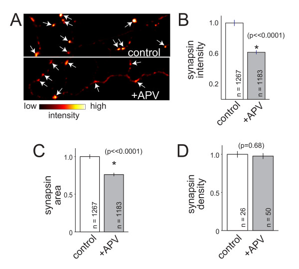
NMDAR-dependent accumulation of synaptic vesicle proteins does not depend on interactions with glia. (A) Images of synapsin immunofluorescence (arrows) at presynaptic terminals of neurons grown in the absence of glia. Images are pseudo-colored such that the highest intensities are white while the lowest are black, as indicated by the intensity scale below the images. Top, control neuron; bottom, APV-treated neuron. (B, C) Synapsin intensity and apparent size at presynaptic terminals were significantly reduced when neurons were treated with APV even when glia were not present. (E) Similar to what was observed in cultures grown in contact with glia, the density of synapsin-positive presynaptic puncta remained unchanged. Data are presented as normalized mean ± standard error of the mean, and the number of puncta measured are indicated on the plot. *, significant difference (P-values are indicated in the figure).

