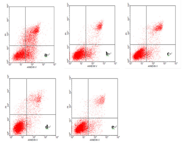
Figure 9.
Effect of PinX1 on nasopharyngeal carcinoma cell apoptosis measured by flow cytometry. Shown are the diagram of flow cytometry of NPC 5-8 F cells stained with Annexin V and propidium iodide solution (PI) and (a) transfected with pEGFP-C3-PinX1, (b) transfected with pEGFP-C3, (c) treated with lipofectamine alone, (d) untreated and (e) t transfected with PinX1-FAM-siRNA, respectively. The upper and lower right quadrants represent apoptotic cells and the lower left quadrant represents normal cells. The data indicate that the number of apoptotic cells transfected with pEGFP-C3-PinX1 was significantly greater than that of cells treated otherwise.

