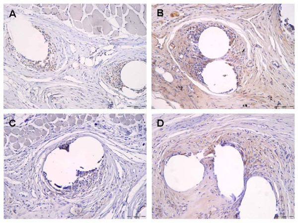
Figure 2.
Immunohistochemical images representing MMP-2 staining following implantation of a PVDF mesh (A) and a PVDF+PAAc+8 μg/mg mesh (C). Figure (B) demonstrates ß-galactosidase staining following implantation of a PVDF mesh and a PVDF+PAAc+8 μg/mg mesh (D) (magnification 200 fold, 90 days postoperatively respectively).

