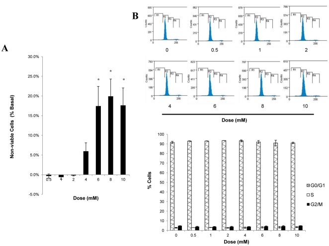
Figure 7. Effect of CGA on cell viability and cell proliferation of L6 myotubes.
Myotubes were incubated with incremental concentrations of CGA for 24 hours. A: Viability of myotubes was measured using MTT staining. Readings are expressed as a percentage of non-viable cells compared to vehicle-treated myotubes. B: Numbers of cells in cell-cycle phases were examined using propidium iodide staining and FACS analysis. Readings are expressed as percentage of cells stained by propidium iodide at different phases. Results are the mean ± SE of three independent experiments. *P<0.05 compared with vehicle-treated control.

