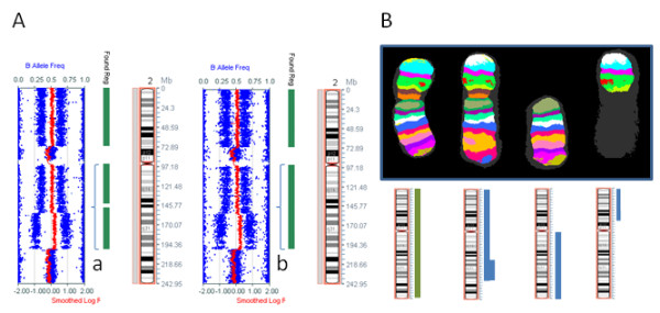
Figure 4.

A comparison of SNP array results for chromosome 2 using DNA from fresh and fixed cells. A. A comparison of SNP array results from fresh (left) and fixed (right) U937 cell line. Gain of a section of the long arm (brackets a, b) is denoted by a single vertical green bar for the fixed specimen (Found Reg = Found Region) (b) whereas in the fresh specimen (a) this is divided into two separate sections. This is representative of the minor boundary differences determined by the Karyostudio software, between the two experiments. B. The M-BAND pattern for chromosome 2. The idiograms below the banded chromosomes show the section of chromosome 2 present in each chromosome. The green bar represents the homolog from one parent and the blue bars represent the homolog from the other parent (inferred from B allele frequencies, see Methods). The Illumina CytoSNP 12 array probes have a median spacing of 6.1 kb.
