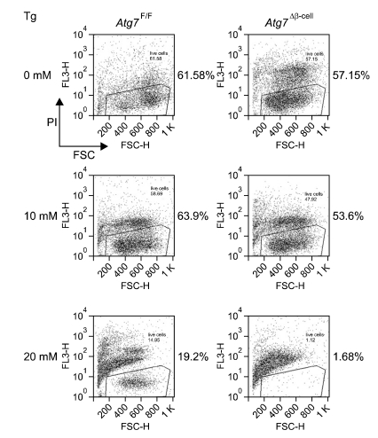
Figure 2.
Viability of autophagy-deficient islet β-cells from Atg7Δβ-cell mice after treatment with thapsigargine (Tg), a classical ER stressor. Viability presented in this figure as the number on each panel was measured by FACS analysis after staining with propidium iodide. Autophagy-deficient β-cells were more susceptible to thapsigargin-induced cell death, probably because of insufficient UPR gene expression.

