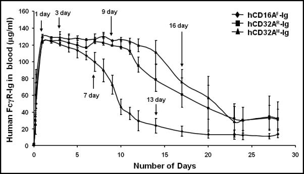
Figure 2. Hydrodynamic based expression of human FcγR-Ig molecules in mice.

Human FcγR-Ig genes in plasmid DNA (10 μg) were diluted in 1.6 ml of sterile saline and injected within 5 seconds into each group of mice through the tail vein. Subsequently, 5 μl blood samples were collected at different time points and diluted in PBS/EDTA. Plasma was separated to detect FcγR-Ig dimers using ELISA as described under Materials and Methods. The plasma from a group of normal mice injected with PBS served as a specificity control. Purified hCD16A-Ig and hCD32A-Ig were used as positive controls, while BSA coated wells served as negative controls. Purified hCD32A-Ig was used as a standard to quantify the level of dimers in the blood. Experiments are representative of three individual experiments. Data are mean ± SD of triplicates.
