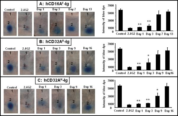
Figure 3. Hydrodynamically expressed human FcγR-Ig molecules block antibody mediated inflammation in mice.

Mice (n=12) were injected with 10 μg of plasmid vector containing human FcγR-Ig cDNA (Panel A: hCD16AF-Ig, Panel B; hCD32AR-Ig, Panel C; hCD32AH-Ig) intravenously as described under Material and Methods. RPA was carried out using three mice at each time point. Mice were injected intradermally with PBS (site 1) and 25 μg (site 2) of anti-Ova per site. RPA was initiated by injecting Ova with 1% Evan's blue intravenously through the tail vein. The antibody (2.4G2) treated control mice were injected with 25 μg/ml of blood (40 μg/mouse) mAb. The PBS injected mice served as the untreated positive control. After 3 h the mice were euthanized, and the dorsal side of the skin was photographed for analysis. The figure is representative of three individual mice. Bar graph on the right side represents the quantitative analysis of RPA. The dermal lesion, seen blue in the photographs, was quantified using ImageJ and KaleidaGraph softwares for groups with or without FcγR dimer treatment. Data are presented as the mean ± SD from three experiments. *p<0.01, **p<0.001.
