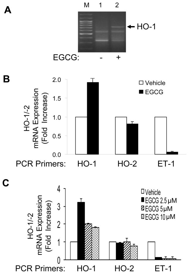
Figure 1.
Treatment of endothelial cells with EGCG increased HO-1 mRNA expression. A. HAEC were serum-deprived for 2 h and then treated with vehicle or EGCG (10 μM, 8 h). Total RNA was isolated from these cells and subjected to differential gene analysis as described in Methods. M = marker lane. B. HAEC were serum-deprived for 1 h and then treated with vehicle or EGCG (10 μM, 8 h). One μg of total RNA was reverse transcribed and subjected to quantitative real-time PCR for HO-1, HO-2, ET-1, and β-actin using QuantiTect SYBR Green PCR with appropriate primer sets as described in Methods. HO-1, HO-2, and ET-1 mRNA expression were normalized to β-actin mRNA expression. Data shown are the mean ± SEM of 4 independent experiments (vehicle vs. EGCG treatment for HO-1, HO-2, and ET-1; p < 0.02, p > 0.18, and p < 0.0005, respectively). C. HAEC were treated as in panel B except various concentrations of EGCG were used as indicated. Total RNA was subjected to quantitative real-time PCR analysis as described in panel B. Data shown are the mean ± SEM of 4 independent experiments. EGCG treatment caused increased expression of HO-1 (but not HO-2) mRNA and attenuated ET-1 mRNA expression (p < 0.001 by ANOVA and Dunnett’s post-test).

