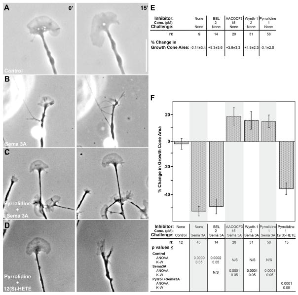
Figure 2.
Effect of PLA2 inhibition on Sema3A-induced growth cone collapse. Growth cones of DRG neurons in explant cultures (on laminin) were exposed by bath application to either vehicle (control), Sema3A, or 12(S)-HETE, with or without pyrrolidine present. A and B, phase contrast micrographs of growth cones before (left panel) or 15 min after (right panel) bath application of either control medium or Sema3A. C and D, growth cones pre-incubated (15 min) with 1μM pyrrolidine, before (left panel) and 15 min after (right panel) bath application of either Sema3A or 10−8M 12(S)-HETE. Scale bar 10μm. E, growth cone area changes (mean % change ± SEM) over 15min in the presence of PLA2 inhibitors, without Sema3A challenge. These values are not significantly different from that for control incubation. F, quantitative analysis of growth cone collapse in the presence of various PLA2 inhibitors. Results are expressed as mean % change in growth cone area ± SEM, measured at t=0 min and t=15 min after onset of challenge. The inhibitor and concentration used, the challenge, and the n values are indicated below each condition, as are the p values when contrasting individual measurements. Because the distribution of values was marginally normal, p values are shown for both parametric (ANOVA, FDR) and non-parametric (K-W, Dunn’s procedure) statistical analyses. The overall p value was <0.0001 for both analyses. Grey shading has been added to facilitate reading of the graph.

