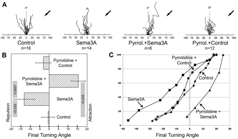
Figure 4.
Effect of GIVA PLA2 inhibition on Sema3A-induced growth cone turning. Growth cones of DRG neurons (explants on laminin) were exposed to gradients of control medium or Sema3A in the presence or absence of 1μM pyrrolidine. A, rosebud plots of axonal responses. Plots illustrate growth cone translocation during gradient exposure for 1 h, shown at a time resolution of 5 min. Arrows mark the position of the micropipette tip. The abscissa indicates distance in micrometers, and the ordinate marks 0° in an arc from −90° to +90°. B, final turning angles (means ± SEM) in response to gradients of either control medium or Sema3A, with or without pyrrolidine present in the culture media. p values are shown in the grey rules whose ends designate the individual measurements to be contrasted (ANOVA, FDR). The overall p value was <0.0009. C, cumulative distribution plots of turning angles in response to (from left to right) Sema3A, Pyrrolidine + Control, Control, and Pyrrolidine + Sema3A. The vertical grey line marks the 0° turning angle.

