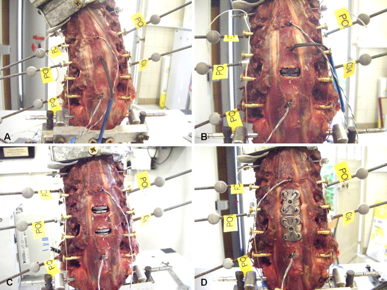
Fig. 2.
Group A: intact (a), 1-level TDR (b), 2-level TDR (c) and 2-level arthrodesis (d). Cervical specimens were mounted on the set up with reflective markers fixed on each vertebra from C3 to C7 to allow for measurement of 3D displacements using an optoelectronic measurement system. Pressure sensors were inserted in adjacent discs to measure IDP during flexibility tests

