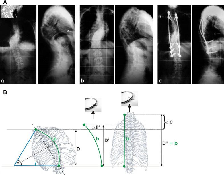
Fig. 4.
A A clinical example of improvement in neurological function during HGT. a A 13-year-old patient with congenital kyphoscoliosis and progressive neurologic deficit (Nurick III, Ranawat IIIa, incontinency, leg weakness), a scoliosis of 63° and a kyphosis of 75°. b After 11 days in HGT, the neurology had improved, as had the kyphosis (slightly). c Treatment with an anterior release T6–8 and T11–L1 and 2-staged PSF T6–L2. At 48 months’ follow-up, the patient was defined as Ranawat II. The patient was satisfied. B Clinical rationale for the improvement of neurologic symptoms during HGT. Rigid kyphoscoliosis can decompensate, and tensioning of the spinal cord being draped over the kyphotic or scoliotic apex might create a myelopathic spinal cord with progressive neurologic deficits. Improvements in progressive neurological deficits in selective cases with decompensated curves can occur during HGT. The effect is induced by a slight curve correction. This, in turn, causes a release of the apical tether on the spinal cord. With partial correction (image in the middle), the tension on the spinal cord is reduced, which allows the cord to recover preoperatively. The clinical observations can be put into an algebraic equation, as illustrated the length of the spinal cord sector can be estimated using the formula:  During HGT, the trunk height increases (D vs. D′), and the kyphotic and scoliotic curves are reduced slightly, but rigid curves do not fully correct, which should be avoided. With slight curve correction, the apical tension on the cord is reduced. If HGT is applied until complete straightening of the spine occurs (see D″), tension-induced myelopathy may result

