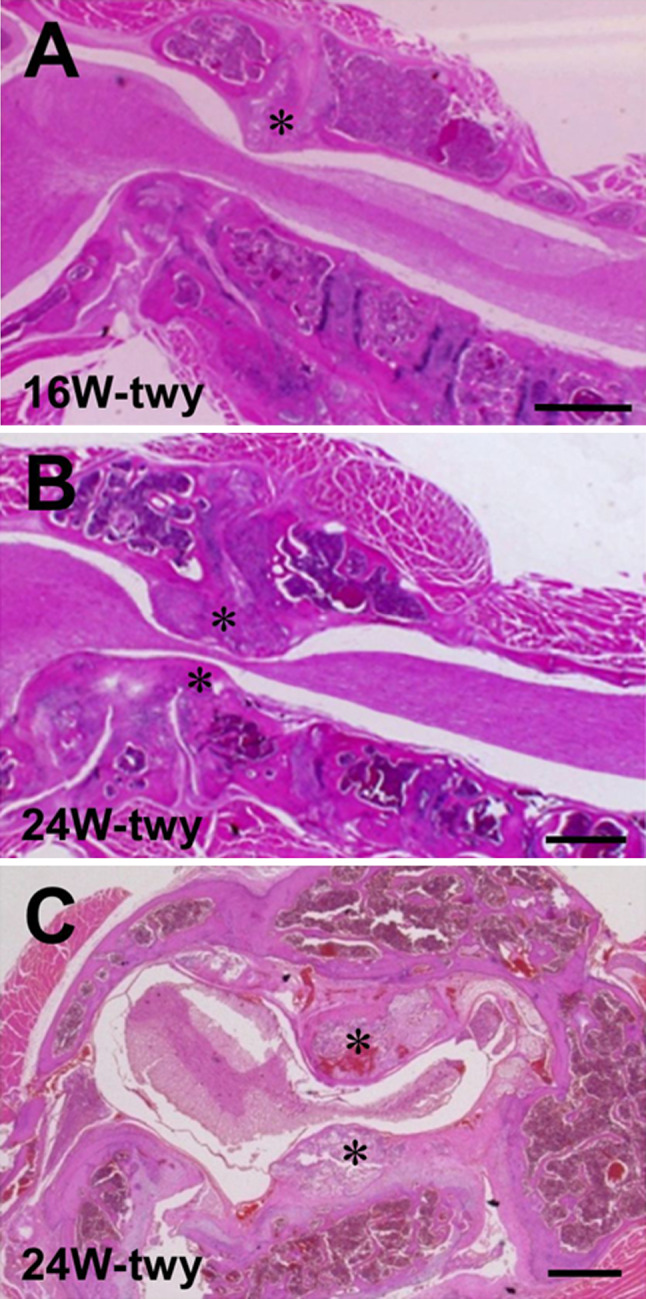
Fig. 1.

Photographs showing representative hematoxylin and eosin (H&E)-stained sagittal (a, b) and transaxial (c) sections of the cervical spine and spinal cord in 16-week-old (a) and 24-week-old twy/twy mice (b, c). Calcified lesions originating from atlantoaxial membrane in twy/twy mice grow progressively with age, compressing the spinal cord between C2 and C3 segments laterally or posteriorly. Asterisk calcified lesions. Scale bar 500 μm (a, b), 200 μm (c)
