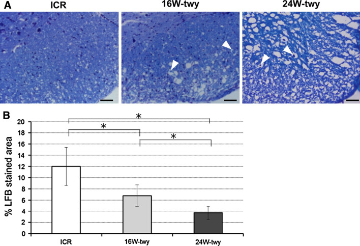
Fig. 2.
Photographs showing representative LFB staining of the ventral microcystic cavity in 16 and 24 weeks twy/twy and ICR mouse. Spotted white holes caused by separation of myelin sheaths from the axons in the anterior and lateral columns in twy/twy mouse. Note also swelling and deformity (arrow heads) of axons particularly in the anterior columns in twy/twy mice (a). The percentage of the cross-sectional area of residual tissue was decrease with age in twy/twy mice and, being significantly smaller when compared with ICR mice (b). Data are mean ± SEM (n = 4). *P < 0.05

