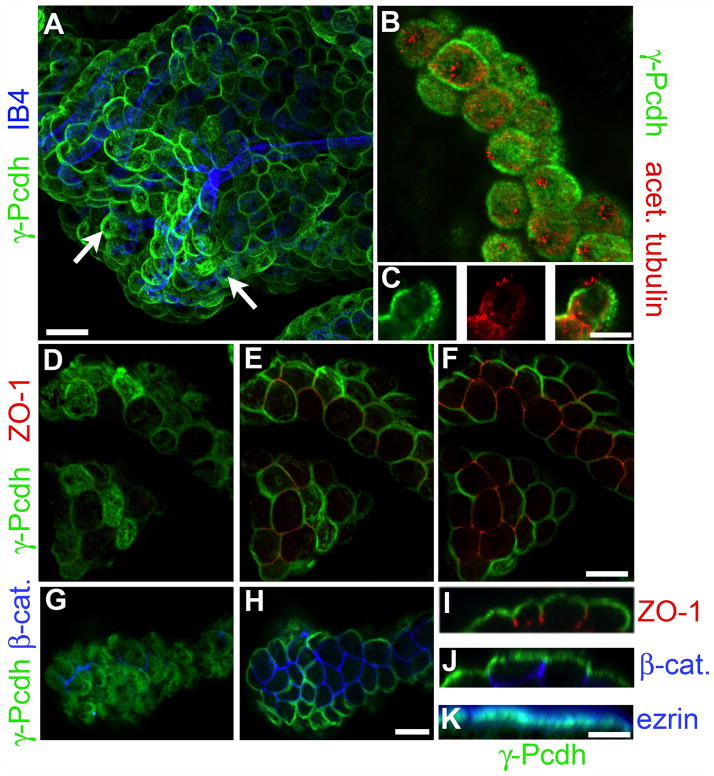
Figure 2. γ-Pcdh proteins are apically localized in CP epithelial cells.
Whole-mount preparations of Pcdh-γfus CP were immunostained with the indicated antibodies or lectins: GFP, for γ-Pcdh-GFP fusion proteins; isolectin B4 (IB4), a blood vessel marker (A); acetylated tubulin, a cilia marker (B,C); ZO-1, a tight junction marker (D–F,I); β-catenin, a basolateral adherens junction marker (G,H,J); and ezrin, an apical sub-membrane marker (K). A and B are maximal projections through a confocal z-stack; D–F and G–H show selected planes within stacks, moving deeper into the tissue. C and I–K show 90° rotated cross-sections through a portion of a z-stack. The γ-Pcdhs are restricted to the apical surface and absent from stromal blood vessels (A). They are also excluded from the patch of membrane from which the cilia protrude (B,C). As expected for an apical protein, there is no overlap with ZO-1 (D–F,I), which marks tight junctions that lie just basal to the apical surface, or with β-catenin, a basolateral marker (G,H,J), but extensive overlap with ezrin (K). Scale bars are 30 μm A, 12μm B–H, and 10μm I–K.

