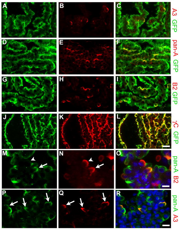
Figure 6. Individual γ-Pcdhs are differentially expressed among choroid plexus epithelial cells.
Cryostat sections of fourth ventricle adult Pcdh-γfus choroid plexus, immunostained using the indicated antibodies in each row of panels; right panel in each row shows a merged image. While GFP antibody, which detects all γ-Pcdhs in the Pcdh-γfus mouse, labels the apical surface of every epithelial cell, monospecific antibodies against A3 (B) or B2 (H) label only a small, apparently random, subset of cells. Pan-A antibody, which recognizes 11 of the 22 γ-Pcdhs, labels a larger subset of the cells (E). As expected, anti-γ-Pcdh constant domain antibody (γC) staining completely overlaps that of GFP (J–L). Double-labeling with the pan-A antibody and anti-A3 (P–R) shows that A3-expressing cells make up a small subset of those cells that express any of the A subfamily proteins. Double-labeling with the pan-A antibody and anti-B2 (M–O) demonstrates that some CP epithelial cells express both B2 and some A subfamily γ-Pcdhs (arrow), while some express B2 in the absence any A subfamily γ-Pcdhs (arrowhead); conversely, many cells that express A subfamily members (green) do not express B2. Together, these results show that different subsets of γ-Pcdh proteins localize to the apical surface in individual CP epithelial cells. Scale bar is 25 μm for (A–L), 12.5 μm for (M–R).

