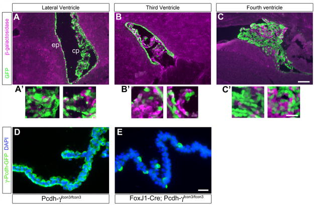
Figure 7. FoxJ1-Cre restricts mutation of the Pcdh-γ gene cluster to the choroid plexus and ependyma within the brain.
A FoxJ1-Cre transgenic line was crossed the Z/EG reporter mouse in order to visualize the spatial pattern of Cre recombinase activity. In Z/EG, β-galactosidase is ubiquitously expressed until Cre excises the β-gal cassette, leading to subsequent expression of EGFP. Widespread Cre activity is detected by GFP staining in the lateral and fourth ventricle choroid plexus and ependyma (A, A′, C, C′); Cre activity is very patchy, however, in the third ventricle choroid plexus (B, B′). Insets (A′–C′) are magnified views of the choroid plexus from each of the panels. Cre activity can also be demonstrated in FoxJ1-Cre; Pcdh-γfcon3/fcon3 choroid plexus as loss of the floxed GFP-tagged constant exon 3 (D, E). The vast majority of choroid plexus epithelial cells are knocked-out in this restricted mutant. Scale bar is 150 μm in A–C, 50 μm in A′–C′, and 30 μm in D–E.

