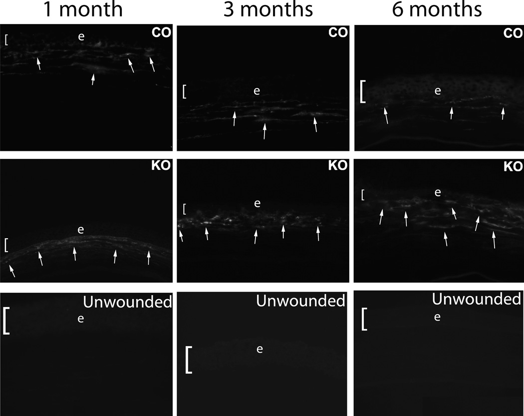
Fig. 2.
Immunohistochemistry for α-smooth muscle actin (SMA) in the central corneal stroma of control (CO) and IL-1 receptor knockout (KO) mouse corneas with haze at one month, three months or six months following irregular PTK. SMA+ myofibroblast density was significantly higher in the knockout than control mice, especially at the six-month time point, where myofibroblast density had markedly declined in the control cornea compared to three-month time point after irregular PTK. In the IL-1 receptor knockout mice, the SMA+ myofibroblast density has not declined and actually appears increased in this individual section. In unwounded corneas, SMA+ cells were not detected in either unwounded IL-1 receptor knockout (unwounded) or unwounded control (not shown) corneas. Representative SMA+ myofibroblasts are indicated by arrows. The bracket and e indicates the corneal epithelium in each panel. Magnifications 400X.

