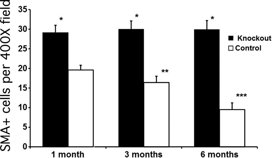
Fig. 3.
Quantitative analysis of α-smooth muscle actin-positive myofibroblasts in central cornea stromal cells/400× microscope field at one month, three months, or six months after irregular PTK in the IL-1 receptor knockout and control groups. Error bars represent SEM. * indicates that the density of SMA+ cells was significantly different (p < 0.001) in the knockout that had irregular PTK compared to the control that had irregular PTK at one month, three months or six months after surgery. SMA+ cell density in the central corneal stroma did not significantly decrease over time in the KO corneas from one month to three months after surgery or from three months to six months after irregular PTK. Conversely, SMA+ myofibroblast density in the central corneal stroma significantly decreased between one and three months (**) or between three and six months (***) after irregular PTK in control corneas.

