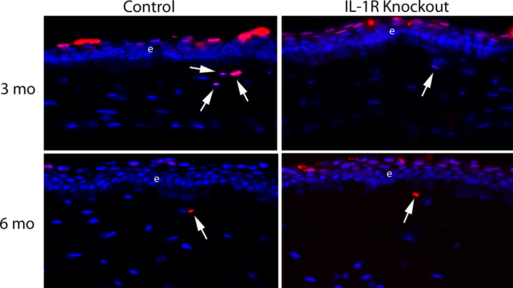
Fig. 4.
Representative TUNEL+ stromal cells (arrows) in the anterior 50 µm of stroma in the IL-1 receptor knockout group (A) and control group (B) at three and six months after irregular PTK. Arrows indicate rare TUNEL+ cells in the anterior 50 µm of stroma in each group. e indicates the epithelium. Note there are also normal TUNEL+ cells in the apical epithelium of each cornea. Magnifications 400X.

