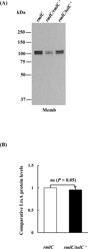
Figure 4. Detection of leukotoxin in the rmlC mutant strain overexpressing TolC.
(A). Immunoblot analysis. Equivalent amounts of outer membrane (OM) protein of the rmlC mutant (rmlC), the rmlC complemented strain (rmlC/rmlC+), and the TolC overexpression strain (rmlC/tolC+) were separated, transferred to PVDF, and probed with anti-LtxA antibody. (B). Quantification of leukotoxin (LtxA). The integrated intensity of each band in panel A, which represents the relative protein level, was quantified using the ImageJ program. The membrane-bound leukotoxin found in the rmlC/tolC+ strain remained the same as the rmlC mutant. The signal intensity of the rmlC mutant was arbitrarily set as 1.0. (ns: nonsignificant)

