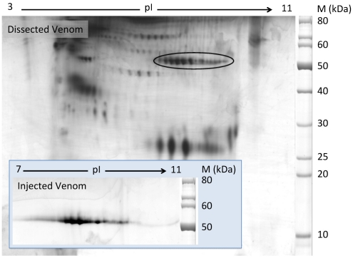
Figure 1.
Two-dimensional gel electrophoresis of C. consors dissected and injectable venoms. IEF was performed under denaturing conditions using a 3–11 NL IPG strip followed by 10% SDS-PAGE in Tris/Taurine buffer in a second dimension. After silver staining and identification by ESI-MS/MS, 10 and 8 Hyal isoforms were identified in DV and IV respectively. All the DV Hyals are located in the encircled region. The IV Hyal spots are shown in the inset at the bottom left.

