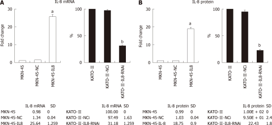
Figure 1.
Interleukin-8 expression of gastric cancer cells. A: Real-time polymerase chain reaction determined the level of Interleukin-8 (IL-8) mRNA in gastric cancer (GC) cells. Expression levels were determined by the ΔΔCt method using glyceraldehyde-3-phosphate dehydrogenase as endogenous control. Histograms show fold change over the relative expression levels of IL-8 mRNA of control cells. Each bar represents the mean ± SD (aP < 0.05 vs MKN-45, bP < 0.05 vs KATO-III); B: IL-8 production in GC cells measured by enzyme-linked immunosorbent assay. Histograms show fold change in the relative expression levels of IL-8 protein of control cells. All experiments were repeated three times with similar results. Each bar represents the mean ± SD (aP < 0.05 vs MKN-45, bP < 0.05 vs KATO-III).

