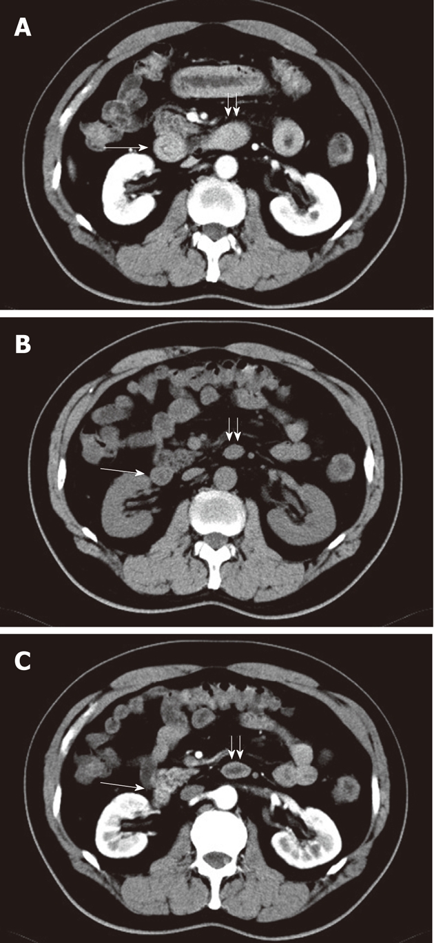
Figure 1.

A 55-year-old man. A: Circumferential thickening of the proximal small bowel in a long segment including the descending duodenum (long arrow) and horizontal duodenum (double short arrows) in venous phase image; B and C: This bowel segment appears normal in unenhanced image (B) and arterial phase image (C).
