
Figure 1. Structural surface representations of MHC class I (A) and MHC class II (B) molecules in complex with peptide shown in stick representation.
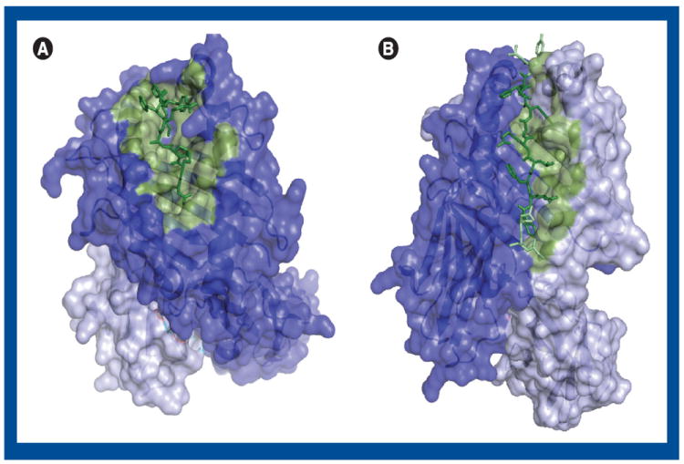
(A) The peptide is green and the residues in the α chain considered important for peptide binding [73] is colored dust green. The rest of the α chain is dark blue and β-2-microglobulin is light blue. (B) The binding core of the peptide is dark green, and the rest of the peptide is bright green. The residues in the β chain considered important for peptide binding [83] are colored dust green. The rest of the β chain is colored dark blue, and the α chain is light blue. The figure has been created using PyMol with the PDB available templates of an HLA-A*11:01-peptide complex structure, 2HN7 [105] (A) and an HLA-DR1-peptide structure, 1AQD [106] (B).