Figure 1.
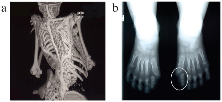
Clinical features of fibrodysplasia ossificans progressiva (FOP). The classic clinical phenotype of FOP is characterized by two features:
(a) The extensive heterotopic bone formation typical of FOP is seen in a three-dimensional reconstructed computed tomography (CT) scan of the back of a twelve-year-old child. Ribbons, sheets, and plates of bone form in extra-skeletal connective tissue and fuse the joints of the axial and appendicular skeleton. For comparison, an image of a normal skeleton (back view) can be viewed at: http://www.shutterstock.com/pic.mhtml?id=11313556.
(b) Radiograph of the feet of a three-year-old child shows symmetrical great toe malformations (a circle indicates the left foot malformation) of metatarsals and proximal phalanges along with microdactyly, fused interphalangeal joints, and hallux valgus deviations at the metatarsophalangeal joints.
[Reprinted from Shore et al., Nature Genetics 2006, 38, 525–527.]

