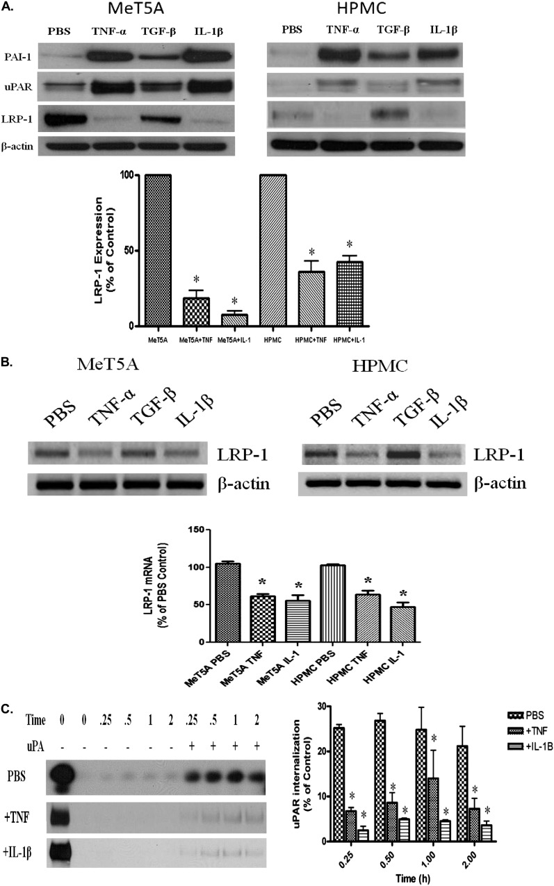
Figure 2.
TNF-α and Il-1β decrease LRP-1 expression in PMCs. (A) Serum-starved MeT5A and primary HPMCs were treated with TNF-α, TGF-β, and IL-1β for 12 hours at 37°C in serum-free RPMI. The cells were then lysed and probed for LRP-1, uPAR, PAI-1, and β-actin. Images are representative of three independent experiments. *P < 0.01. (B) Serum-starved MeT5A and HPMCs were treated with TNF-α, TGF-β, and IL-1β as described in Methods. RNA was isolated from the cells, transcribed into cDNA, and probed for LRP-1 cDNA. β-actin cDNA was used as a loading control. (C) Serum-starved MeT5A cells were treated with PBS, TNF-α, or IL-1β for 12 hours. The cells were then biotinylated and incubated in the presence or absence of uPA for 0, 0.25, 0.50, 1, and 2 hours. The cells were subjected to glutathione (GSH) washes, lysed, and probed for uPAR internalization. Images are representative of three independent experiments.

