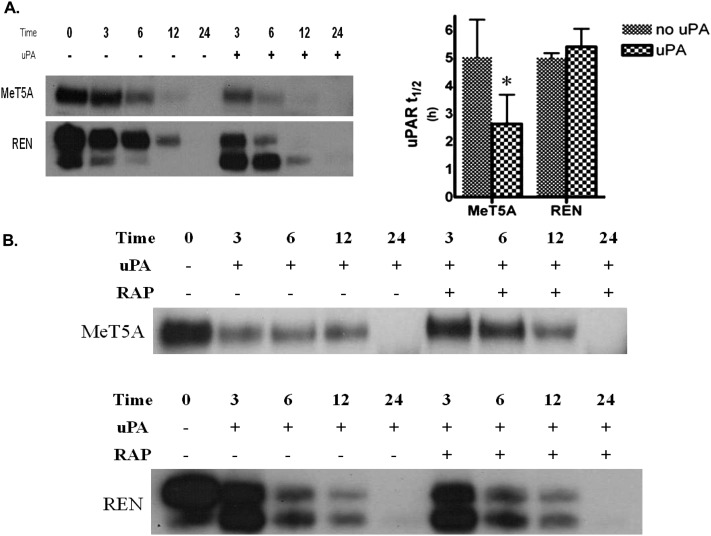
Figure 4.
uPAR t1/2 measurements in PMC and LRP-1–deficient MPM cells. (A) Serum-starved MeT5A and REN cells were surface biotinylated and treated with 20 nM uPA for 0, 3, 6, 12, and 24 hours in SFM. Biotinylated proteins were then isolated from lysates, resolved on SDS-PAGE, and probed for uPAR. The images are representative of at least three independent experiments. (B) Serum-starved MeT5A and REN cells were treated with uPA in the presence and absence of 200 nM RAP. The t1/2 of biotinylated uPAR protein was calculated via densitometric analysis, and the calculated t1/2 for the control and uPA treated samples were statistically analyzed. The data presented represent the average of three independent experiments. *P < 0.05.

