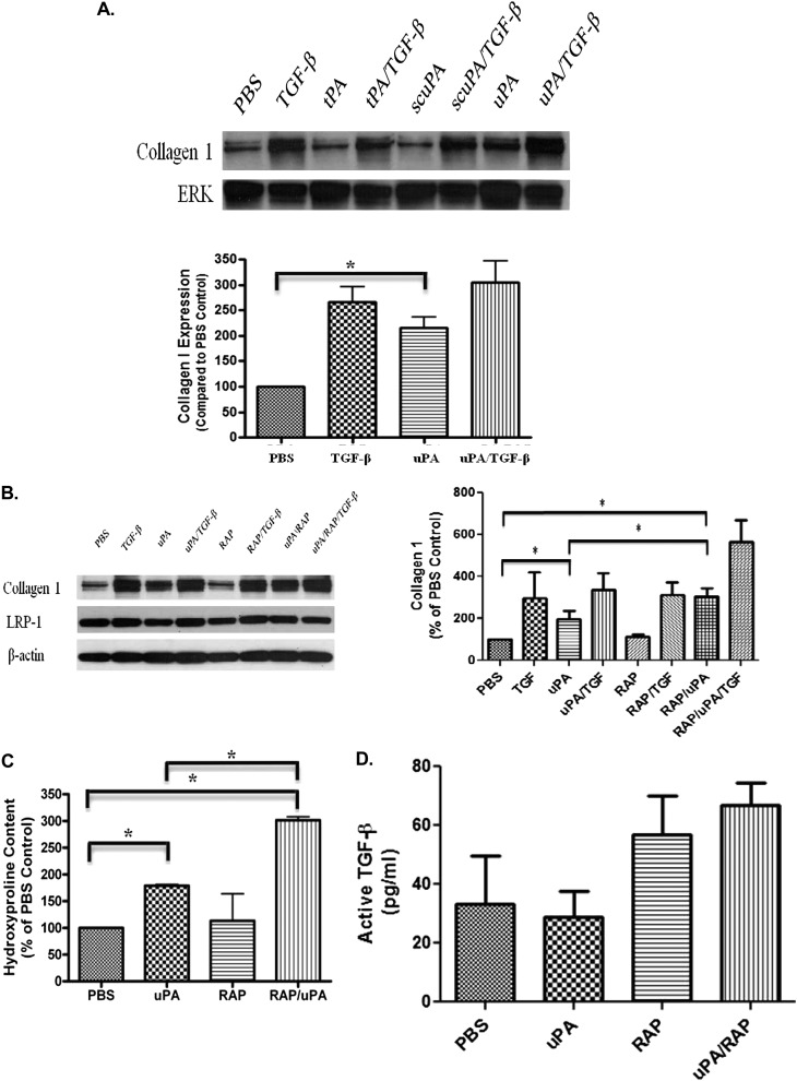
Figure 7.
uPA increases collagen 1 expression in primary PMCs. (A) Primary RPMCs were treated with tPA (tissue type plasminogen activator; 20 nM), scuPA (single chain or proenzyme uPA; 20 nM), and uPA (two chain uPA; 20 nM) in the presence and absence of TGF-β (5 ng/ml). Conditioned media and lysates were collected, resolved on SDS-PAGE, and probed for collagen 1 and ERK. The images are representative of three independent experiments. (B) HPMCs were treated with uPA and/or TGF-β in the presence or absence of RAP (200 nM) for 48 hours in SFM. Conditioned media and cell lysates were collected, resolved by SDS-PAGE, and subjected to Western blotting for Collagen 1 and LRP-1. β-actin was used in loading controls. The images are representative of three independent experiments. (C) HPMCs were treated with uPA and/or TGF-β in the presence or absence of RAP for 48 hours in SFM. Conditioned media were collected and assayed for hydroxyproline content. This experiment was repeated twice (n = 3 determinations per group in each experiment). *P < 0.05. A representative experiment is shown. (D) HPMCs were treated with uPA in the presence or absence of RAP for 48 hours in SFM. Conditioned media were collected and assayed for active TGF-β via ELISA.

