Fig. 6.
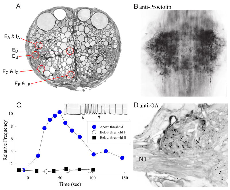
Swimmeret command neurons. (A) Cross-section of the intersegmental connective showing the locations of excitatory and inhibitory swimmeret command neurons (Acevedo, 1990; Acevedo et al., 1994; Wiersma and Ikeda, 1964). Dorsal is at the top. Colored circles show the five locations at which these axons occur. EA & IA: excitatory and inhibitory axons at Wiersma and Ikeda's position A. EB: excitatory axon at position B. EC & IC: excitatory and inhibitory axons at C. ED: excitatory axon at D. EE & IE: excitatory and inhibitory axons at E. (B) Whole mount of a ganglion A2, viewed from the dorsal side with anterior at the top, labeled with anti-proctolin antibody (Acevedo et al., 1994). Many processes and puncta in each LN are densely labeled. (C) Stimulation of certain excitatory command neurons both excites the swimmeret system and releases proctolin that can be recovered from the fluid bathing the nerve cord (Acevedo, 1990). The plot compares the proctolin recovered during a bout of above-threshold stimulation with that recovered during two bouts of below-threshold stimulation of the same command axon. Proctolin was detected using the frequency of spontaneous movements of a detached locust leg as a bioassay (O'Shea and Adams, 1981). The inset shows the increased frequency of movements caused by the above-threshold sample; arrows mark application and removal of the sample from the leg. (D) Cross-section through the left LN and the base of N1 of an abdominal ganglion labeled with anti-octopamine antibodies (Eckert et al., 1992; Schneider et al., 1993). The midline is to the right and dorsal is at the top. Within the LN, many puncta and fine processes are densely labeled.

