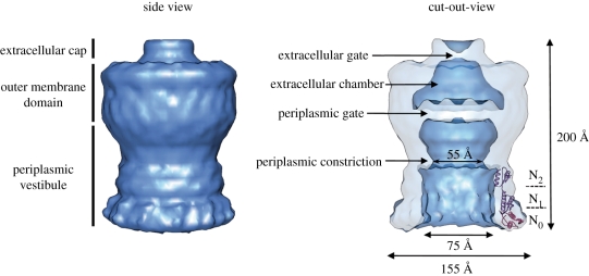
Figure 3.
Electron microscopy structure of T2SS secretin. Cryo-EM reconstitution of V. cholerae T2SS secretin GspDQ at 19 Å resolution (EMDB1763 and adapted from [61] by permission from Nature Publishing Group). The GspDQ cryo-EM density reveals a cylindrical channel assembly 155 Å in diameter and 200 Å in length. In side view, three domains are identified from top to bottom: the extracellular cap, the outer-membrane domain and the periplasmic vestibule domain. In a cutout view, secretin contains an extracellular chamber limited by an extracellular gate and a periplasmic gate. The vestibule domain shows a constriction which results in a narrowing of the channel diameter from 75 to 55 Å. The crystal structure of the N-terminal periplasmic subdomains N0–N1–N2 from ETEC [62] is fitted into the GspDQ periplasmic vestibule (adapted from [47] by permission from Elsevier).

