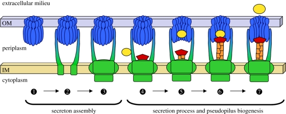
Figure 5.
Model of secreton assembly and operation. Schematic of secreton biogenesis as discussed in this review. The first part of the secreton to be assembled is proposed to be the secretin  . The next step could be the successive recruitment of the trans-periplasmic protein GspCP
 and of the inner membrane surface
  . The recognition of the substrate by the T2SS takes place in the periplasm and may involve a peripheral element of the secreton, GspCP
 . The substrate is then transferred to the secretin vestibule
  in which it could contact the pseudopilus tip complex that is emerging from the inner membrane surface
  . The exoprotein could then be released in the extracellular medium through the secretin pore
  . The secretin and periplasmic domain of GspCP are shown in blue, the components of the inner membrane surface are shown in green, the pseudopilus and the secreted proteins are shown in orange/red and yellow, respectively.

