Figure 3.
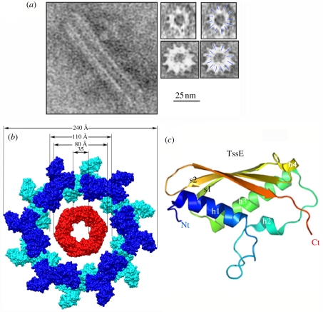
Structure of tube sheath and hub proteins. (a) Electron microscopy side-view (left) or cross section (right) of the TssB/TssC tubes formed in vitro (with permission from Bönemann et al. [41]). The 25 mm scale bar applies to all the electron microscopy views. (b) Molecular surface model of a cross section of a T6SS tube with Hcp rings in the middle (red) and TssB/TssC sheath around (blue, modelled from phage T4 tail sheath [1FOH] [42]). (c) Ribbon model (rainbow colours) of the hub protein TssE, generated from the structure of Geobacter sulfurreducens gp25 (2IA7; Joint Center for Structural Genomics 2006, unpublished data). Figures were drawn with PyMOL [24] or Chimera [25].

