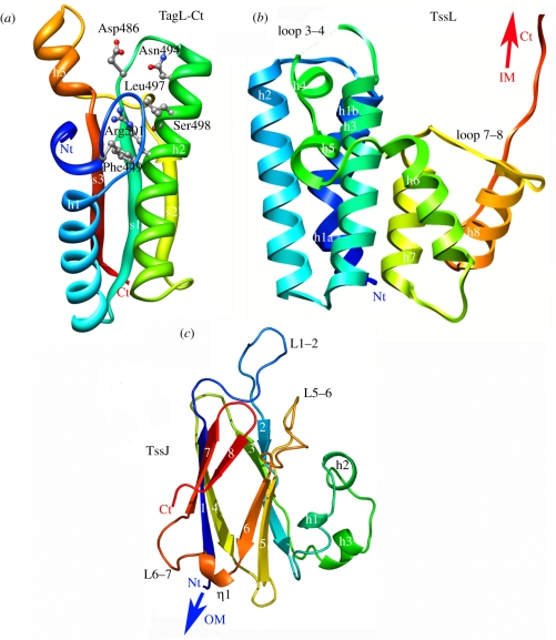
Figure 5.
Structures of the membrane complex proteins. (a) Ribbon view (rainbow colours from blue Nt to red Ct) of the C-terminal peptidoglycan-binding domain of the enteroaggregative E. coli TagL protein (residues 414–557) modelled from the X-ray structure of E. coli YiaD (2K1S; T. A. Ramelot & M. A. Kennedy 2008, unpublished data). (b) Ribbon view (rainbow colours from blue Nt to red Ct) of the N-terminal cytoplasmic domain of the TssL protein from enteroaggregative E. coli [61]. (c) Ribbon view (rainbow colours from blue Nt to red Ct) of the C-terminal periplasmic domain of the TssJ lipoprotein from enteroaggregative E. coli [55].

