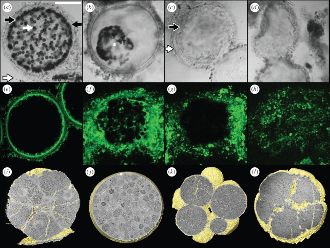
Figure 1.
(a–h) Decay in the giant sulphur bacterium Thiomargarita namibiensis and (i–l) comparison with Doushantuo microfossils. (a–d) Differential interference contrast (DIC) microscopy images of Thiomargarita. (a) Living Thiomargarita cell with diffuse sulphur vesicles surrounding the central vacuole (black arrow, mucous sheath; white arrow, sulphur vesicle; white-lined black arrow, laminar sheets; black-lined white arrow, microbial process). (b) Cell showing sulphur vesicles (region around asterisk) coalescing on the surface of the vacuole. (c) Internal contents have decayed but cell wall (white-lined black arrow) and mucous sheath (black-lined white arrow) remain. (d) Chain of cells where only the distorted mucous sheath remains. (e–h) Confocal microscopy of Thiomargarita. (e) Living cell showing the large central vacuole (image courtesy of V. Salman, Max Planck Institute, Bremen). (f) Cell showing clustered vesicles on the surface of the vacuole. (g) The same cell as in (f), showing the vacuole. (h) Decayed cell showing diffuse organic material. (i–l) Synchrotron radiation X-ray tomographic microscopy images of Tianzhushania specimens from the Ediacaran Doushantuo formation. (i) Specimen showing putative nuclei (NRM-PZ X 4469); (j) single-celled specimen showing putative lipid droplets (NRM-PZ X 4470); (k) cell morphology, but not internal structure, is preserved (NRM-PZ X 4471); (l) only surface morphology is preserved (NRM-PZ X 4472). Scale bars: (a) 100 µm; (b) 95 µm; (c,d) 120 µm; (e) 60 µm; (f,g) 25 µm; (h) 65 µm; (i) 180 µm; (j) 160 µm; (k) 170 µm; (l) 115 µm.

