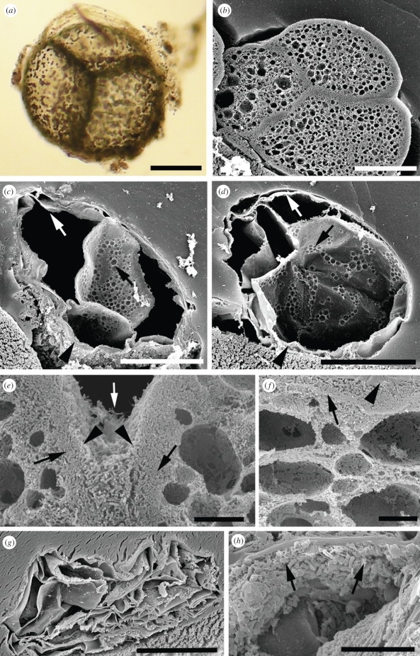
Figure 2.
Taphonomic comparison of Thiomargarita with marine invertebrate embryos. (a) Light micrograph of multi-cellular reductive division-stage Thiomargarita fixed with 2.5% glutaraldehyde. External gelatinous sheath, outer cell wall, internal cell boundaries and intracellular sulphur inclusions are visible. (b–h) Sections viewed by scanning electron microscopy. (b) Cleavage stage embryo of the sea urchin H. erythrogramma, fixed, settled on a Millipore filter and embedded in agarose. Outer fertilization envelope has contracted down onto the embryo; outer cell boundaries, internal cell boundaries, denser cortical cytoplasm and cavities of characteristic lipid droplet vesicles scattered through the internal cytoplasm are visible. (c,d) Reductive division stage Thiomargarita fixed with 2.5% glutaraldehyde at time of collection, then settled on a Millipore filter and embedded in agarose. Unlike embryos, Thiomargarita specimens collapsed. In Thiomargarita sections, external sheaths (arrowheads) are visible at bottom edges; external lamina are present under the sheath (white arrows); internal cell boundaries and sites of intracellular sulphur inclusions (black arrows) are visible. Note the vast central vacuole (black cavity). In (b–d), the Millipore filter substrate is a rough texture at lower left of the panels and embedding agarose is a smooth texture surrounding the specimen. (e,f) Microbial taphonomy of H. erythrogramma embryos. BME-stabilized embryos were incubated with Psuedoalteromanas tunicata, a marine gamma proteobacterium, for 6 days and then fixed in glutaraldehyde. (e) Internal views of a pseudomorphed two-cell embryo sectioned after fixing. The dense surface biofilm (arrowheads) and microbially replaced intracellular fabric (arrows) are visible. The white arrow indicates the pseudomorphed cell boundary between the two embryonic cells. (f) Pseudomorphed embryo embedded before sectioning, as in (b–d). Arrowhead indicates the boundary between embryo surface biofilm and the agarose; arrow shows exterior surface of the embryo. The embryo is partially compressed in the vertical plane by the embedding procedure, as indicated by the slightly flattened lipid vesicle droplets. (g,h) Microbial taphonomy of Thiomargarita stabilized in BME at the time of collection, incubated for 6 days with P. tunicata under the same conditions as in (e,f), then embedded and sectioned as in (b,c,d,f). (g) Microbial-treated Thiomargarita cell. Bacteria do not fill the vacuole, and the specimen is partly collapsed. (h) Enlarged view of part of another microbial-treated Thiomargarita cell. Bacteria do not fill the internal spaces, but partial bacterial biofilms have formed on some of the surfaces of the laminar sheets; arrows indicate bacterial bodies in the biofilm. In (g), the Millipore filter substrate is visible at the bottom right. Scale bars: (a–d,g) 100 µm; (e,f,h) 10 µm.

