
Figure 1. NG2+ cells divide in corpus callosum and exhibit limited self-renewal and oligodendrocyte differentiation ex vivo.
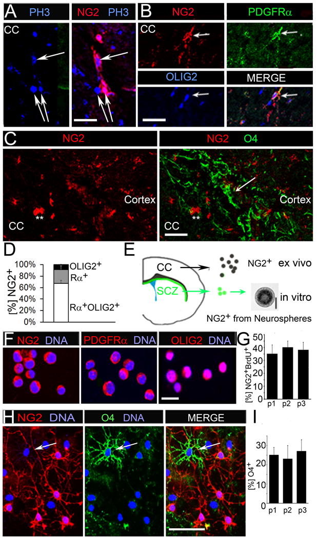
(A) Immunofluorescent co-staining for NG2 and mitosis marker phospho-histone3 (PH3) showing proliferating NG2+ cells in the corpus callosum (CC) of wild type P60 mice.
(B) NG2+ cells co-express OPC markers PDGFRα and OLIG2.
(C) Immunofluorescent co-staining for NG2 (asterisks) and O4 expression (white arrows).
(D) Quantification of OPC marker expression of NG2+ cells in the adult CC. NG2+ cells are PDGFRα/OLIG2/NG2 triple-positive (Rα/OLIG2/NG2; 65%); Rα/NG2 (23%), NG2/OLIG2 (8%) and NG2/O4 (4%) double-positive. 2% of NG2+ cells are OPC marker-negative.
(E) Schematic of a hemisphere of a coronal brain section. NG2+ cells were acutely isolated from CC tissue and also obtained from subcallosal zone (SCZ)-derived neurospheres.
(F) Immunofluorescent staining for NG2, PDGFRα and OLIG2 on acutely isolated NG2+ cells.
(G) BrdU was added to FACS sorted, acutely isolated NG2+ OPC cells after they have grown in culture for 48 hrs, and flow cytometry was used to determine the percentage of NG2 and BrdU double-positive (NG2+BrdU+) cells.
(H) NG2+ cells from p1-p3 were immunostained for NG2 and O4 after ten days in differentiation medium.
(I) Percentage of O4+ cells remained 23-26% for three passages. Scale bars in (A) and (F) 10μM, (B) 50 μM, (C), (E) and (H) 100μM. Error bars are +/− standard error of the mean (SEM). See also Figure S1.