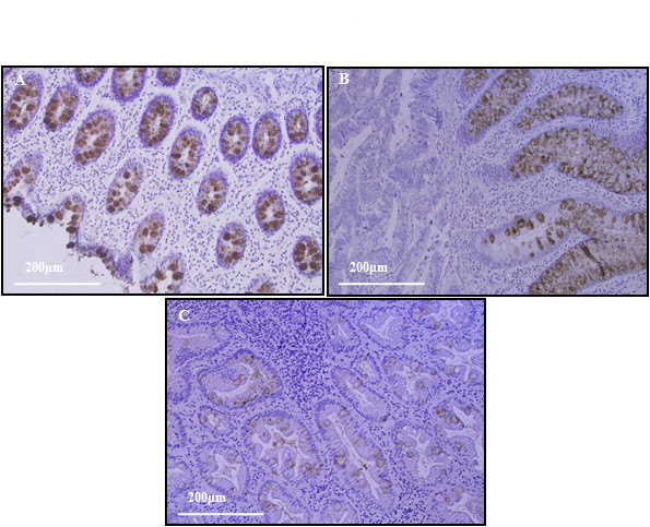
Figure 6.

Immunohistochemistry analysis of IgGFcBP expression. (A) Healthy colon tissue with a specific IgGFcBP staining localized in the intracellular mucus of globelet cells. (B) CRC tissue with a positive IgGFcBP staining present in the non-invasive zone. (C) Mixed hyperplastic/adenomatous polyp with the IgGFcBP positive glands in the non dysplastic areas.
