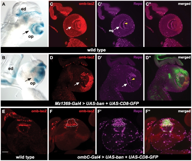
Figure 5. bantam down regulates omb in the optic lobe.
(A, B) The brains are positioned for lateral views. X-gal staining indicates omb-lacZ expression patterns in the optic lobe. eye disc (ed), optic lobe (op). (A) wild type brains; (B) over expression of bantam by the optic lobe driver, Mz1369-Gal4. (C–D″) A single focal plane of lateral view. UAS-CD8-GFP (green) is used to view expression of Gal4. Anti-β-galactosidase (red) is to view expression of omb-lacZ. Glia cells are viewed by anti-Repo (megenta). (C–C″) wild type; (D–D″) over expression of bantam and UAS-CD8-GFP by the optic lobe driver, Mz1369-Gal4. (C–C″) and (D–D″) are at a similar focal plane. Medulla glia cells are indicated by white arrows and medulla neuropile glial cells are indicated by yellow arrow heads. (E–F″) A single focal plane of a horizontal view is shown. (E) wild type brains; (F–F″) over expression of bantam and UAS-CD8-GFP by ombC-Gal4. Increased glial cells are noted by a dashed line. Scale bar: 50 µm.

