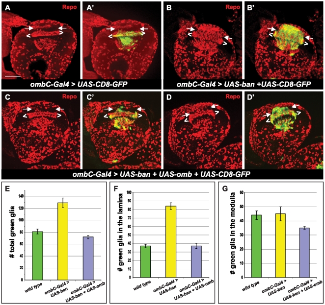
Figure 6. omb rescues bantam.
All brains are positioned for a horizontal view. All pictures are maximum projections from multiple sections. Anti-Repo staining (red) is used to label glial cells. ombC-Gal4 expression is visualized by UAS-CD8-GFP (green). Glial cells located between white brackets (< >) correspond to the three layers of laminal glial cells: epithelial, marginal and medulla glia. Glial cells in the lamina are located between two arrows. Genotypes: (A, A′) UAS-CD8-GFP/+; ombC-Gal4/+; (B, B′) UAS-CD8-GFP/+; ombC-Gal4/UAS-ban; (C–D′) UAS-omb/UAS-CD8-GFP; ombC-Gal4/UAS-ban. (E–G) Histograms of green glia number in the optic lobe of the third instar larvae. (E) total green glia in the optic lobe; (F) the green glia in the lamina; (G) the green glia in the medulla. Scale bar: 50 µm.

