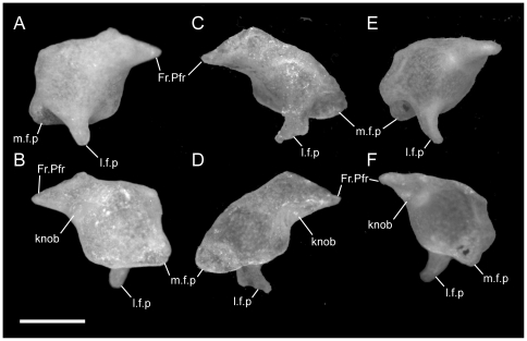
Figure 13. Disarticulated prefrontals.
Anterior is to the left in A,D,E; anterior is to the right in B,C,F; scale bar = 0.5 mm. A–C from the left side of the skull in U. woodmasoni (TMM M-10001); D–F from the right side of the skull in U. melanogaster (TMM M-10045); and G–I from the left side of the skull in B. rhodogaster (TMM M-10027). A,C,E in lateral view and B,D,F in medial view. Fr.Pfr = frontal process of prefrontal; knob = medially projecting knob at base of frontal process of prefrontal; l.f.p = lateral foot plate; m.f.p = medial foot process.

