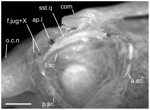
Figure 26. Magnified view of the posterior end of the crista circumfenestralis of Uropeltis woodmasoni (TMM M-10008) in dorsolateral view.
Anterior is to the right; scale bar = 0.5 mm. This specimen has a double opening for the passage of cranial nerve X, the jugular vein, and associated tissue. a.sc = anterior semi-circular canal; ap.l = apertura lateralis recessus scalae tympani; com = compound bone; f.jug = jugular foramen; l.sc = lateral semi-circular canal; o.c.n = neck of occipital condyle; p.sc = posterior semi-circular canal; sst.q = suprastapedial process of the quadrate; X = foramen for vagus nerve.

