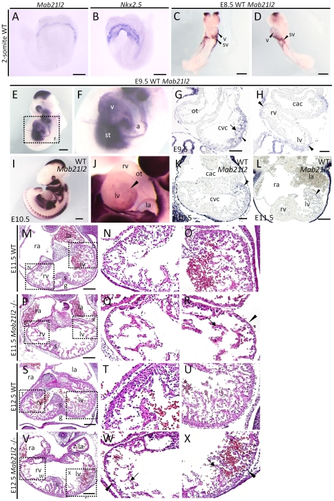
Figure 1. Mab21l2 is required for the formation of the trabecular and compact myocardium.
(A–F) Whole-mount in situ hybridization of 2-somite, E8.5 and E9.5 wild-type (WT) embryos for the indicated transcripts. (A, B) At the 2-somite stage, Mab21l2 appeared to be expressed adjacent to the first heart field expressing Nkx2.5. (C and D) Expression of Mab21l2 was detected in the ventricle, and sinus venosus at E8.5. (E and F) Mab21l2 was expressed in the ventricle (v) and the septum transversum mesenchyme (st) at E9.5. Scale bar represents 60 µm(A, B) and 300 µm (C–E). (G and H) In situ hybridization analysis of Mab21l2 in transverse paraffin sections of E9.5 WT embryonic heart. (G) Mab21l2 is expressed in the trabecular myocardium (arrow) and the compact myocardium (arrowhead). (H) Mab21l2 is expressed in the right and left ventricle (black arrowheads). Scale bars represent 50 µm (G) and 30 µm (H). (I and J) Whole-mount in situ hybridization of E10.5 WT embryos for Mab21l2. (J) Lateral view of the heart shown in (I). Scale bar in I represents 500 µm. (K and L) In situ hybridization analysis for Mab21l2 in transverse paraffin sections of E10.5 (K) and E11.5 (L) WT embryonic hearts. (K and L) Expression of Mab21l2 is seen on the left side of the left ventricle (arrowheads). Scale bars represent 50 µm in (K) and 100 µm (L). (M–X) Hematoxylin and eosin (H&E)-stained transverse sections of WT and Mab21l2-mutant embryonic hearts at E11.5 and E12.5. At E11.5, Mab21l2 mutant embryos show defects in the left ventricle (R), but not in the right ventricle (Q), including reduced trabecular myocardium (arrow) and thin compact myocardium (arrowheads) compared with WT embryos (N and O). Defects were observed in the right (W) and left (X) ventricle in Mab21l2-mutants compared with WT embryos at E12.5 (T and U) (compact myocardium, arrowheads; trabecular myocardium, arrows). a, atrium; cac, common atrium chamber; cvc, common ventricular chamber; ot, outflow tract; g, ventricular groove; la, left atrium; lv, left ventricle; ra, right atrium; rv, right ventricle; v, ventricle; s, interventricular septum; st, septum transversum mesenchyme; sv, sinus venosus. Scale bar represents 100 µm (M, P, S and V).

